cell body reorganization in the spinal cord after sympathectomy

The amount of compensatory sweating depends on the patient, the damage that the white rami communicans incurs, and the amount of cell body reorganization in the spinal cord after surgery.
Other potential complications include inadequate resection of the ganglia, gustatory sweating, pneumothorax, cardiac dysfunction, post-operative pain, and finally Horner’s syndrome secondary to resection of the stellate ganglion.
www.ubcmj.com/pdf/ubcmj_2_1_2010_24-29.pdf

After severing the cervical sympathetic trunk, the cells of the cervical sympathetic ganglion undergo transneuronic degeneration
After severing the sympathetic trunk, the cells of its origin undergo complete disintegration within a year.

http://onlinelibrary.wiley.com/doi/10.1111/j.1439-0442.1967.tb00255.x/abstract

Thursday, June 27, 2013

Sympathectomy (ETS or ESB) can alleviate socia phobia and common fears such as fear of flying, heights, open spaces, or the darkness

Is sympathectomy the new lobotomy?

"ESB may also alleviate social phobia and common fears such as fear of flying, heights, open spaces, or the darkness. In addition, it can be used to decrease trembling of the body, hands, and voice, even stuttering. It may help in alcoholism or drug withdrawal, because these are often linked with social anxiety. 
Sympathetic block is a gentle and exact endoscopic procedure. It is performed as day surgery under light anesthesia."
http://www.sympatix.fi/?lang=en

Friday, January 11, 2013

Patients complain of emotional blunting following endoscopic thoracic sympathectomy for hyperhidrosis and blushing


Hohmann, G.W. : The effect of dysfunction of the autonomic nervous system on experienced feelings and emotions
Paper read at Conference on Emotions and Feelings at New School for Social Research, New York, Oct 1962.
Sherrington, C.S.: Experiments on the value of vascular and visceral factors for the genesis of emotion 
Proc. Roy.Soc., 1900, 66, 390-403
Wenger, M.A.: Emotion as visceral action: an extension of Lange's theory. In Reymert, M.L., Feelings and emotions: the Moosehart ymposium, New York: McGraw-Hill, 1950. Pp.3-10.
Wynne, L.C., and Solomon, R.L.: Traumatic avoidance learning: acquisition and extinction in dogs deprived of normal peripheral autonomic function. 
Genet. psychol. Monogr., 1955, 52, 241-84
Landis, C., and Hunt, W. A. Adrenalin and emotion. 
Psychol. Rev., 1932, 39, 467-85.
Schachter, S., and Wheeler, L.: Epinephrine, chlorpromazine, and amusement. 
J. of abnorm. soc. Psychol., 1962, 65, 121-28.

Wednesday, January 2, 2013

in the absence of autonomic arousal, behavior that appears emotional will not be experienced as emotional


"In the presence of a barking dog, for example, the sympathectomized cats manifested almost all of the signs of feline rage. Finally, Cannon notes the report of Dana (1921) that a patient with a spinal-cord lesion and almost totally without visceral sensation still manifested emotionality.
For either the Jamesian or the present formulation such data are crucial, since both views demand visceral arousal as a necessary condition for emotional arousal. When faced with this evidence, James's defenders (e.g., Wenger, 1950; Mandler, 1962) have consistently made the point that the apparently emotional behavior manifested by sympathectomizied animals and men is well-learned behavior, acquired long before sympathectomy. There is a dual implication in this position: first, that sympathetic arousal facilitates the acquisition of emotional behavior, and second, that sympathectomized subjects act but do not feel emotional. There is a small but growing evidence supporting these contentions. Wynne and Solomon (1955) have demonstrated that sympathectomized dogs acquire an avoidance response considerably more slowly than control dogs. Further, on extinction trials most of their 13 sympathectomized animals extinguished quickly, whereas not a single one of the 30 control dogs gave any indication of extinction over 200 trials. Of particular interest are two dogs who were sympathectomized after they had acquired the avoidance response. On extinction trials these two animals behaved precisely like the control dogs - giving no indication of extinction. Thus, when deprived of visceral innervation, animals are quite slow in acquiring emotionally-linked avoidance responses and in general, quick to extinguish such responses." (p. 163)

"A line of thought stimulated by the Wynne and Solomon (1955) and the Hohmann (1962) studies may indeed be the answer to Cannon's observations that there can be emotional behavior without visceral activity. From the evidence of these studies, it would appear, first, that autonomic arousal greatly facilitates the acquisition of emotional behavior but it is not necessary for its maintenance if the behavior is acquired prior to sympathectomy; and second, that in the absence of autonomic arousal, behavior that appears emotional will not be experienced as emotional." (p. 167)

Psychobiological Approaches to Social Behavior

P. Herbert LeidermanDavid ShapiroHarvard Medical School. Dept. of PsychiatryUnited States. Office of Naval Research - 1964 - 203 pages

Monday, December 10, 2012

The vasodilating effect of spinal dorsal column stimulation is mediated by sympathetic nerves - Springer

The vasodilating effect of spinal dorsal column stimulation is mediated by sympathetic nerves - Springer: "Immediately after sympathectomy, the contralateral right arm became increasingly cold and cyanotic and the patient complained of chronic painful coldness and severe cold-intolerance in the right arm. Attempts to pharmacologically vasodilate the arm with felodipine did not affect the local vasoconstriction and pain. Dorsal column stimulation (associated with symmetrical paraesthesia in both arms) induced an immediate and marked (ten-fold) increase in skin blood flow in the right arm (and in the leg), whereas skin blood flow in the left arm remained unaffected. The lack of vasomotor response in the left arm was not due to maximal vasodilatation at rest, since skin blood flow in the left arm showed a normal capacity for axon reflex vasodilatation following antidromic activation of sensory afferents. The results suggest that the marked vasodilatation induced by dorsal column stimulation is mediated by sympathetic vasomotor fibres, via modulation of central neuronal circuits involved in the regulation of skin sympathetic discharge."

'via Blog this'

Saturday, December 1, 2012

Transection below T8-T10 is not accompanied by reflex sweating

Reflex sweating in patients with spina... [Arch Phys Med Rehabil. 1977] - PubMed - NCBI: "Sweat glands derive their innervation from the sympathetic nervous system. The spinal sympathetic structures that are located in the intermediolateral areas extend from T1-L2 segments and are under the control of hypothalamic centers. Cord transection abolishes the supraspinal control of sudorimotor function. Since sympathetic innervation does not follow a clear segmental distribution, normal sweating may be preserved at a higher or lower level than skin sensation. Nonthermoregulatory reflex sweating is an indication of unchecked spinal cord facilitation and is precipitated by afferent stimuli from bladder, rectum, and various other sources. It is usually a manifestation of mass reflex or autonomic crisis and occurs particularly in cervical or high thoracic lesions. Transection below T8-T10 is not accompanied by reflex sweating. The phenomenon of thermal relfex sweating is controversial. Although some aspects of nonthermoregulatory reflex sweating are still unclear, proper immediate and continuing preventive management will reduce the incidence of this and other autonomic manifestations. "

'via Blog this'

Monday, November 26, 2012

Hemodynamic changes in vertebral and carotid arteries were observed after sympathicotomy for hyperhidrosis


T3 sympathicotomy segment was the most frequent transection done (95.83%), as only ablation (25%) or in association with T4 (62.50%) or with T2 (8.33%). It was observed increase in RI and PI of the common carotid artery (p < 0.05). The DPV of internal carotid artery decreased in both sides (p < 0.05). The SPV and the DPV of the right and left vertebral arteries also increased (p < 0.05). Asymmetric findings were observed so that, arteries of the right side were the most frequently affected.

CONCLUSIONS: Hemodynamic changes in vertebral and carotid arteries were observed after sympathicotomy for PH. SPV was the most often altered parameter, mostly in the right side arteries, meaning significant asymmetric changes in carotid and vertebral vessels. Therefore, the research findings deserve further investigations to observe if they have clinical inferences.
http://www.ncbi.nlm.nih.gov/pubmed/16186983 

Thursday, October 25, 2012

Research indicates that a highly variable heart rate increases your capacity to respond and adapt to life’s challenges.

Research has shown that reductions in heart rate variability are a predictor of sudden cardiac death, even in individuals without a prior history of cardiovascular disease.

http://theconversation.edu.au/depression-can-break-your-heart-literally-1102

Monday, October 22, 2012

Unexpected beneficial effect of stellate ganglion block in a schizophrenic patient

Unexpected beneficial effect of stella... [Can J Anaesth. 2002 Aug-Sep] - PubMed - NCBI: "Unexpected beneficial effect of stellate ganglion block in a schizophrenic patient"

Stellate ganglion block may relieve hot flashes by interrupting the sympathetic nervous system

Stellate ganglion block may relieve hot flash... [Med Hypotheses. 2007] - PubMed - NCBI: "the wide range of conditions that have been reported to respond favorably to stellate ganglion block suggest that its effectiveness may not be solely the result of increased blood flow nor restricted just to its sphere of innervation. We have found that stellate ganglion block is effective in the treatment of hot flashes in postmenopausal women, as well as those with estrogen depletion resulting from breast cancer treatment. Based on evidence that hot flashes may be centrally mediated and that the stellate ganglion has links with the central nervous system nuclei that modulate body temperature, we hypothesize that the stellate ganglion block provides relief of hot flashes by interrupting the central nervous system connections with the sympathetic nervous system, allowing the body's temperature-regulating mechanisms to reset. If this mechanism can be confirmed, this would provide women with intractable hot flashes with an effective, potentially long-lasting means of relieving their symptoms, and potentially widen the range of indications for stellate ganglion block to include other centrally mediated syndromes."

'via Blog this'

Thursday, October 18, 2012

These observations further emphasize our ignorance of the mechanisms responsible for primary hyperhidrosis and of the effect of sympathetic ablation


"These observations further emphasize our ignorance of the mechanisms responsible for primary hyperhidrosis and of the effect of sympathetic ablation on the function of the remaining sympathetic system."  

"Only investigators who deviate from accepted standards innovate and thus advance science. Obviously, such deviations may also result in disasters;"  

Statement made by the former President of the International Society of Sympathetic Surgery,  and ETS surgeon, Moshe Hashmonai (Invited Commentary)   Endoscopic Lumbar Sympathectomy Following Thoracic Sympathectomy in Patients with Palmoplantar Hyperhidrosis    

World J Surg (2011) 35:54–55 DOI 10.1007/s00268-010-0809-5

Monday, September 24, 2012

Post-sympathectomy pain

Postsympathectomy limb pain, postsympathectomy parotid pain, and Raeder's paratrigeminal syndrome are pain states associated with the loss of sympathetic fibres and in particular with postganglionic sympathetic lesions. There is a characteristic interval of about 10 days between surgical sympathectomy and onset of pain. It is proposed that this pain in man is correlated with the delayed rise in sensory neuropeptides seen in rodents after sympathectomy. These chemical changes probably reflect the sprouting of sensory fibres and may result from the greater availability of nerve growth factor after sympathectomy. The balance between the sensory and sympathetic innervations of a peripheral organ may be determined by competition for a limited supply of nerve growth factor.
Lancet. 1985 Nov 23;2(8465):1158-60
http://www.ncbi.nlm.nih.gov/pubmed/2414615?dopt=Abstract

A significant admission from an ETS surgeon, President of the ISSS:

"General represents the ESB (whether as ETS as ETSC or ELS) a substantial interference in regulatory processes of the body represent the decision for this operation requires that previously conservative treatments were made. An ESB is therefore at the end of a treatment history, and never at the beginning."
http://www.dhhz.de/index....;subPage=&section=32

it is an automatic translation (google) from german, so you have to look at what the 'gist' of what he is saying: ETS, ESB = "a substantial interference in regulatory processes of the body"

Friday, August 31, 2012

Wednesday, August 15, 2012

"sympathectomy highlighted the disparity between what is known in practice and what appears in the literature"


The March 2004 edition was quite outstanding, with an excellent editorial reminding the reader that only good results are published. The review on thoracoscopic sympathectomy highlighted the disparity between what is known in practice and what appears in the literature. 
‘Know Your Results’, the topic of the ASGBI Annual Scientific Meeting, is of outstanding importance; what is more, the surgeon has to go on knowing his/her results to ensure standards of practice do not slip.
The Journal appreciates comments and criticism and the correspondence column remains a crucial part of the BJS in its interaction between editors and reader. It is also part of the scientific process.
A more robust and incisive criticism of articles known to be flawed would prevent the retractions that have recently been published in the Lancet.
Christopher Russell, Chairman, BJS Society
Association of Surgeons of Great Britain and Ireland, ANNUAL REPORT 2004

Monday, August 13, 2012

sympathectomy induced morphological alterations in the masseter muscles


Sympathectomized animals showed varying degrees of metabolic and morphological alterations, especially 18 months after sympathectomy. The first five groups showed a higher frequency of type I fibres, whilst the oldest group showed a higher frequency of type IIb fibres. In the oldest group, a significant variation in fibre diameter was observed. Many fibres showed small diameter, atrophy, hypertrophy, splitting, and necrosis. Areas with fibrosis were observed. Thus cervical sympathectomy induced morphological alterations in the masseter muscles. These alterations were, in part, similar to both denervation and myopathy. These findings indicate that sympathetic innervation contributes to the maintenance of the morphological and metabolic features of masseter muscle fibres.

Sunday, August 12, 2012

Bilateral cervical sympathectomies should be avoided because of the destruction of cardioaccelerator tone

http://www.hiesiger.com/physicians/physicianrfl.html

SURGICAL SYMPATHECTOMY ON THE SENSITIVITY TO EPINEPHRINE OF THE BLOOD VESSELS OF MUSCULAR SEGMENTS OF THE LIMBS

Pursuing this study of the effect of epinephrine on muscle blood flow, Duff and Swan (10) reported that during intravenous epinephrine infusions the initial marked dilatation was succeeded by a second phase of moderate dilatation in normal but not in sympathectomized limbs. Because of its absence in chronically sympathectomized limbs this secondary vasodilatation was at that time thought to be an indirect vasomotor effect mediated by the sympathetic nerves. Re-examination of their data in the light of some subsequent critical experiments now reveals that the difference which they found between normal and sympathectomized limbs may be ascribed largely to vascular hypersensitivity in the later.
In the present paper these additional data are reported, and are incorporated with those of Duff and Swan(10); the whole material being interpreted to provide evidence that hypersensitivity of the vessels of skeletal muscle in the upper and lower limbs may result from pre- and postganglionic sympathectomy in man.
EFFECT OF SURGICAL SYMPATHECTOMY ON THE SENSITIVITY TO EPINEPHRINE OF THE BLOOD VESSELS OF MUSCULAR SEGMENTS OF THE LIMBS, ROBERT S. DUFF
J Clin Invest. 1953 September; 32(9): 851–857.
http://www.ncbi.nlm.nih.gov/pmc/articles/PMC438413/

Tuesday, June 12, 2012

Effects of lumbar sympathectomy: abolition of ejaculation, causalgia in 41, general morbidity of 0.6% and mortality rate of 0.6 %, arrhythmias, cardiac decompensation

Goldstein, reviewing the complications of 791 patients undergoing open lumbar sympathectomy reports 10% of specific complications, such as abolition of ejaculation in 22 patients and causalgia in 41, general morbidity of 0.6% (TEP and bleeding) and mortality rate of 0.6 % (arrhythmias, cardiac decompensation or bleeding) 15.

SIMPACTECTOMIA RETROPERITONEOSCÓPICA FOR TREATMENT OF LUMBAR plantar hyperhidrosis RETROPERITONEOSCOPIC lumbar sympathectomy FOR THE TREATMENT OF PLANT HYPERIDROSIS
Marcelo de Paula Loureiro, TCBC-PR ¹, ² Neomar Roman, Sheila Cristina Weigmann ³;
Aline ³ Fontana, Paulo Cesar Bufara Boscardim4
(Rev Bras ECR. Cir. 2007, 34 (4): 222-224).
(translated by google)

dynamic cerebral autoregulation is altered by ganglion blockade

We measured arterial pressure and cerebral blood flow (CBF) velocity in 12 healthy subjects (aged 29+/-6 years) before and after ganglion blockade with trimethaphan. CBF velocity was measured in the middle cerebral artery using transcranial Doppler. The magnitude of spontaneous changes in mean blood pressure and CBF velocity were quantified by spectral analysis. The transfer function gain, phase, and coherence between these variables were estimated to quantify dynamic cerebral autoregulation. After ganglion blockade, systolic and pulse pressure decreased significantly by 13% and 26%, respectively. CBF velocity decreased by 6% (P <0.05). In the very low frequency range (0.02 to 0.07 Hz), mean blood pressure variability decreased significantly (by 82%), while CBF velocity variability persisted. Thus, transfer function gain increased by 81%. In addition, the phase lead of CBF velocity to arterial pressure diminished. These changes in transfer function gain and phase persisted despite restoration of arterial pressure by infusion of phenylephrine and normalization of mean blood pressure variability by oscillatory lower body negative pressure.
Conclusions-: These data suggest that dynamic cerebral autoregulation is altered by ganglion blockade. We speculate that autonomic neural control of the cerebral circulation is tonically active and likely plays a significant role in the regulation of beat-to-beat CBF in humans.
Circulation. 106(14):1814-1820, October 1, 2002.
http://www.problemsinanes.com/pt/re/dyslipidaemia/abstract.00003017-200210010-00017.htm;jsessionid=PX6phQHYFG5PD1p2DMS1cJLvG1TbtLLLH0bfJT6vKJgLLx1zn0Xf!1816077220!181195629!8091!-1?nav=reference

Tuesday, June 5, 2012

effect of bilateral cervical sympathetic ganglionectomy on the architecture of pial arteries

The influence of the cranial sympathetic nerves on the architecture of pial arteries in normo- and hypertension was examined. For this purpose the effect of bilateral superior cervical ganglionectomy was evaluated in normotensive rats (WKY) and stroke-prone spontaneously hypertensive rats (SHRSP). The operations were performed at the age of 1 wk, which is just prior to the onset of ganglionic transmission. The length of the inner media contour was measured and the media cross-sectional area was determined planimetrically, with computerized digitalization of projected photographic images of transversely sectioned pial arteries. Four wk after sympathectomy there was a 20% reduction in media cross-sectional area and a consequent reduction in the ratio between media area and calculated luminal radius in the major pial arteries at the base of the brain in WKY but not in SHRSP. Conversely, in small pial arteries linear regression analysis showed that in WKY subjected to ganglionectomy the relationship between media cross-sectional area and luminal radius was significantly larger in arteries with a radius less than 21 microns compared to untreated WKY. No such effect was seen in the corresponding SHRSP vessels. In addition, the cross-sectional area of the internal elastic membrane (IEM) in the basilar arteries of WKY was measured by means of a computerized image-analysing system. Mean cross-sectional area of the IEM was approximately 45% larger following SE than in control animals. The present findings propose a 'trophic' role for the sympathetic perivascular nerves in large pial arteries of the rat. The increased media-radius ratio in the small pial arteries of the WKY following sympathectomy might reflect a compensatory hypertrophy due to reduced protection from the larger arteries against the pressure load. The inability to detect any morphometrically measurable effect of the sympathectomy in the cerebral arteries of SHRSP is probably explained by a marked growth-stimulating effect of the high pressure load in these animals.
http://www.ncbi.nlm.nih.gov/pubmed/7701941

Tuesday, May 29, 2012

significant fall in left circumflex coronary flow was proportional to the decline in external heart work due to sympathectomy

http://www.springerlink.com/content/k2n6j4555g16x773/

sympathectomy affects the heart, sweating, and circulation

heart rate was significantly reduced at rest (14%), at sub-maximal exercise (12.3%), and at peak exercise (5.7%), together with a significant increase in oxygen pulse (11.8, 12.7, and 7.8%, respectively). The rate pressure product (RPP) was also significantly reduced following the surgical procedure at all three study stages, while all other physiological variables measured remained unchanged. It is suggested that thoracic-sympathetic denervation affects the heart, sweating, and circulation of the respective denervated region

Eur J Appl Physiol. 2008 Sep;104(1):79-86. Epub 2008 Jun 10.

Sunday, May 20, 2012

Drug warning - Karvezide, AVAPRO HCT - 'you must tell your doctor if you have had sympathectomy'

Tell your doctor if:

* you have had a sympathectomy

* you have been taking diuretics

*you have a history of allergy or asthma


www.racgp.org.au/cmi/swckarvz.pdf


2. Before you start to take AVAPRO HCT

Tell your doctor if:
  • you suffer from any medical conditions especially-
    - kidney problems, or have had a kidney transplant or dialysis
    - heart problems
    - liver problems, or have had liver problems in the past
    - diabetes
    - gout or have had gout in the past
    - lupus erythematosus
    - high or low levels of potassium or sodium or other electrolytes in your blood
    - primary aldosteronism
  • you are strictly restricting your salt intake
  • you are lactose intolerant or have had any allergies to any other medicine or any other substances, such as foods, preservatives or dyes.
  • have had a sympathectomy
  • you have been taking diuretics
  • you have a history of allergy or asthma
http://www.mydr.com.au/medicines/cmis/avapro-hct-300-25-tablets

Published by MIMS/myDr March 2011
UBM Medica Australia uses its best endeavours to ensure that at the time of publishing, as indicated on the publishing date for each resource (e.g. Published by MIMS/myDr January 2007), the CMI provided was complete to the best of UBM Medica Australia's knowledge.  

Thursday, May 17, 2012

Use of stellate ganglion block for the treatment of psychiatric and behavioral disorders

The present invention is directed to a method for the treatment of a patient suffering from psychiatric and behavioral disorders, including post partum depression, post traumatic stress disorder, compulsive smoking, attention deficit hyperactivity disorder, gambling addiction, comprising the step of administering a stellate ganglion block to the patient to alleviate the symptoms. The stellate ganglion block may be followed by a sympathectomy to provide permanent relief.


Kind Code: A1
http://www.freepatentsonline.com/y2007/0135871.html

Saturday, May 5, 2012

nerves that sent blood-pressure-raising flight-or-fight signals to the brain were cut

page 187:
It was a grueling operation called sympathectomy, in which the nerves that sent blood-pressure-raising flight-or-fight signals to the brain were cut...The nerve cutting scrambled signals to her circulatory system. She was cold on one side of her body and warm on the other.

The Happy Bottom Riding Club: The Life and Times of Pancho Barnes (Paperback)

by Lauren Kessler (Author)

Wednesday, April 25, 2012

Sympathectomy increased the pain threshold and made the sympathectomized rats hypesthetic

Normal adult rats were sympathectomized at L2-L3. The threshold for thermal noxious pain by hot-plate analgesia test and changes in neuropeptides in the lumbar dura mater and dorsal root ganglia using light microscopic immunohistochemistry were assessed and compared with control rats.
Results: In the hot-plate analgesia test, sympathectomized rats increased their hot-plate latency time compared with that of sham-operated rats. Density of calcitonin gene-related peptide immunoreactive fibers in sympathectomy side of the lumbar dura mater decreased to 45.5% compared with the contralateral side. The number and size of calcitonin gene-related peptide immunoreactive cells in dorsal root ganglia showed no difference between sympathectomized and contralateral side.
Conclusion: Sympathectomy increased the pain threshold and made the sympathectomized rats hypesthetic. A large numbers of sensory fibers innervated the lumbar dura mater via L2-L3 sympathetic nerve in rats. Sympathectomy reduced the number of these nerve fibers in the lumbar dura mater. Sympathetic nerves may play an important role for low back pain involving the lumbar dura mater.
http://journals.lww.com/spinejournal/Abstract/1996/04150/An_Anatomic_Study_of_Neuropeptide.4.aspx

Long-term sympathectomy induces sensory and parasympathetic fibres sprouting, and mast cell activation in the rat dura mater

http://discovery.ucl.ac.uk/1330488/

There are similarities between the delayed onset of the human pain state and the delayed rise in sensory peptides after sympathectomy

The effect of sympathectomy on the calcitonin gene-related peptide (CGRP) level in the rat primary trigeminal sensory neurone was investigated. Six weeks after bilateral removal of the superior cervical ganglion there was a 70% rise in the CGRP content of the iris and the pial arteries, a 34% rise in the concentration in the trigeminal ganglion but no change in the brainstem. The CGRP rise in both end organs suggests that this phenomenon may be common to all peripheral organs receiving combined sensory and sympathetic innervations. The lack of any rise in the brainstem CGRP content raises the possibility that this process spares central terminations. In contrast, the level of neuropeptide Y, a peptide mainly contained in sympathetic terminals, fell to 35% of control values in the iris and pial arteries whilst the trigeminal ganglion and brainstem concentrations remained unchanged. The possible relevance of these observations to the clinical syndrome of postsympathectomy pain (sympathalgia) is discussed. There are similarities between the delayed onset of the human pain state and the delayed rise in sensory peptides after sympathectomy.
http://www.ncbi.nlm.nih.gov/pubmed/3877546

sympathectomy resulted in complete disappearance of histochemically detected adrenergic and a considerable decrease of cholinergic nerve fibers in the pial arterial walls

sympathectomy resulted in complete disappearance of histochemically detected adrenergic and a considerable decrease of cholinergic nerve fibers in the pial arterial walls. The vasodilatation was much less obvious in sympathectomized than in control animals. This was associated with (and probably caused by) a considerable rise in histochemically detected serotonin activity of the pial arteries walls. After recovery of blood supply to the brain the constriction of the pial arterial active segments restricting the excessive cerebral blood flow, was significantly reduced due, probably, to the sympathetic deprivation. Therefore, the sympathetic control plays an important part in pial arterial responses regulating the adequate blood supply of the cerebral cortex.
http://www.ncbi.nlm.nih.gov/pubmed/7173422

Monday, April 23, 2012

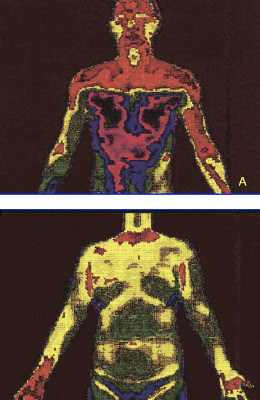
Digital infrared thermal image after T2 sympathicotomy or T3 ramicotomy.

(A) Clear cut change of skin temperature after a T2 sympathicotomy. (B) An even distribution of skin temperature after ramicotomy.
Gossot and colleagues [8] analyzed a group of T2, T3, T4 sympathectomy patients in comparison with a group of patients undergoing a T2, T3, T4 ramicotomy and they reported no statistical difference regarding the incidence of CS between the two groups studied (72.2% and 70.9%). However in terms of the severity of CS (embarrassing, disabling) causing inconveniences to daily life, they reported 27% and 13% incidences in these two groups, respectively. These findings suggest that by preserving the sympathetic trunk, it was possible to reduce the severity of CS.
The preganglionic fibers of the sympathetic nerve to the arm originate mostly from the spinal segments T3–T6 and the postganglionic fibers of the sympathetic nerve to the arm originate from T2 and, to a lesser extent, the T3 ganglia [9]. This implies that the division of preganglionic fibers (rami communicantes) reduces the extent of denervation of the sympathetic nerve as compared with the division of postganglionic fibers (sympathetic trunk) in the treatment of palmar hyperhidrosis.
Sympathectomy or sympathicotomy is one of the procedures used to divide the sympathetic trunk. Sympathicotomy distinctively changes sympathetic nerve distribution in comparison with a ramicotomy. Figure 4A illustrates the clear-cut changes of skin temperature after a T2 sympathicotomy. However the overall sympathetic nerve distribution to the body is not markedly changed after a T3 ramicotomy because a T3 ramicotomy is a procedure that is used to divide one of the preganglionic fibers and to preserve the sympathetic trunk. Figure 4B illustrates an even distribution of skin temperature after T3 ramicotomy.
 http://ats.ctsnetjournals.org/cgi/content/full/78/3/1052#FIG4

Tuesday, April 10, 2012

most surgeons do not have a clear understanding of their short-term outcomes for the majority of procedures they perform

The public would probably be surprised to know that most surgeons do not have a clear understanding of their short-term outcomes for the majority of procedures they perform.

Of even greater concern is the lack of data on long-term outcomes associated with surgical interventions.

Many surgeons argue that they are too busy and do not have the time and resources to conduct this sort of follow-up. This is not entirely without foundation, but it does seem difficult to defend a stance that says “I will continue to work feverishly at the operations I do but not assess how successful my results are”.

Guy Maddern (ASERNIP-s): No excuse for poor surgical outcomes

MJA INSIGHT, 8 August 2011

Monday, March 12, 2012

It’s not unusual to hear people who have undergone sympathectomies describe themselves as feeling emotionally “colder” than before

It’s not unusual to hear people who have undergone sympathectomies describe themselves as feeling emotionally “colder” than before. Among psychologists and neurologists alike there is concern, but no evidence, that the procedure limits alertness and arousal as well as fear, and might affect memory, empathy and mental performance. Professor Ronald Rapee, the director of the Centre of Emotional Health at Sydney’s Macquarie University, says he’s counselled several people who complain of feeling “robot-like” in the long-term wake of the operation. “They’re happy they no longer blush, but they miss the highs and lows they used to feel.”
(John van Tiggelen, Good Weekend Magazine, The Age and the Sydney Morning Herald, 10th March 2012)

Friday, February 24, 2012

impairment of the CBF autoregulation after unilateral cervical sympathectomy

Although these findings argued against a neurogenic mechanism, James at al. (1969) reported impairment of autoregulation after unilateral cervical sympathectomy in the babbon. Gotoh et al. (1971/1972) observed impairment of autoregulation in patients with the Shy-Drager syndrome.
It was concluded that the autonomic nervous system plays an important role in the mechanism of autoregulation of CBF and that his mechanism is independent of the chemical control of the cerebral vessels. This was confirmed by direct observation of the pial vessels in cats, where separate sites of action in the vascular tree for autoregulation and chemical control were demonstrated; the autoregulatory reaction was located in pial arteries with a diameter larger than 50 μ, and the reaction to carbon dioxide in pial arteries of smaller diameter (Gotoh et al. 1975).
They concluded that the arteries operating in autoregulation were the larger ones with the dense innervation, while the smaller arteries with sparse innervation were involved in chemical control.
Coronna and Plum (1973) demonstrated the absence of CBF autoregulation in a patient with a Shy-Drager syndrome who had a postganglionic denervation.

Gotoh et al (1979) subsequently showed that autoregulation in patients with this syndrome was impaired irrespective of the localization of the damage to the cervical sympathetic nervous system (preganglionic, central, postganglionic) as judged by the eye instillation test.
Handbook of Clinical Neurology,

Vascular Diseases, Part I by P. J. Vinken, G. W. Bruyn, H. L. Klawans, and J. F. Toole
, Volume 53, Part 1
Elsevier Health Sciences, 1988

Friday, February 17, 2012

reduced oxygen saturation and shallow respiration after a thoracoscopic sympathectomy

  1. D. J. Canty1,2,3,* and  C. F. Royse4,5
1Department of Anaesthesia, Royal Hobart Hospital, 48 Liverpool Street, Hobart, Tasmania 7000, Australia
  1. 2Medical School of The University of Tasmania, Tasmania, Australia
  2. 3Department of Pharmacology, The University of Melbourne, Melbourne, Australia
  3. 4Anaesthesia and Pain Management Unit, Department of Pharmacology, University of Melbourne, Melbourne, Australia
  4. 5Royal Melbourne Hospital, Victoria, Australia
  1. *Corresponding author. E-mail: david.canty@dhhs.tas.gov.au 
  2.     http://bja.oxfordjournals.org/content/103/3/352.full

CAUSES AND MANAGEMENT OF ORTHODEOXIA - The Australian Short Course on Intensive Care Medicine, 2005

DEFINE AND LIST THE CAUSES AND MANAGEMENT OF PLATYPNOEA AND
ORTHODEOXIA

p. 79:
Autonomic
o Parkinson disease (Hussain 2004)
o Bilateral thoracic sympathectomy (van Heerdon 2004)

Published in 2005 by
The Australasian Academy of Critical Care Medicine
“Ulimaroa”
630 St Kilda Rd, Melbourne,
Victoria 3004

ISSN 1327-4759

HAZARDS ASSOCIATED WITH CERVICO-THORACIC SYMPATHECTOMY

The following is a case report of a healthy 18-year-old woman who had bilateral Cervico- Thoracic sympathectomy done in two stages for severe hyperhidrosis in the palms of her hands.
Two episodes of asystolic arrest occurred during the 2nd stage left Cervico-Thoracic sympathec- tomy.

Thirty-five minutes after starting the operation, as the surgeon was retracting and dissecting the upper thoracic chain,
the cardiac monitor showed sudden onset of sinus bradycardia. The pulse rate was 50 beats per minute. Atropine 1·2 mg was given intravenously but cardiac asystole occurred.

External cardiac compression was started and another dose of atropine 1· 2 mg was given, followed by adrenaline 1·0 mg but there was no response. Following a second dose of adrenaline 1·0 mg and sodium bicarbonate 100 mEq, the
heart restarted with a marked sinus tachycardia.

The cause of hyperhidrosis apparently originates from some poorly understood stimulation of the sympathetic nervous system (Cloward 1969), and in sensitive patients this may possibly lead to excessive vagal stimulation to counteract it, as illustrated by the bradycardia and asystolic reaction to the sudden removal of the sympathetic control, and by the high doses of sympathomimetic drugs necessary to recommence cardiac activity. Anatomically the heart is innervated by the cardiac plexus which consists of the cardiac nerves derived from the cervical and upper thoracic ganglia of the sympathetic trunk and branches of the vagus.The pacemaker of the heart, the sino-atrial node, is innervated by both the parasympathetic and sympathetic nerves (King and Coakley 1958). The ventricular muscle of the heart is supplied solely by the sympathetic nerves, and the larger branches of the coronary arteries are also predominantly innervated by sympathetics (Woollard 1926). These factors may also have a bearing on the hazard of a bilateral cervico- thoracic sympathectomy, which leaves the heart solely under vagal control. Usually, following
denervation, the heart will initiate its own impulse, without recourse to external agencies, but there may be a place for transvenous electrode cardiac pacing, if spontaneous initiationof impulse is delayed, or bradycardia is severe.


R. F. Y. ZEE*
Royal Perth Hospital, Perth
Anaesthesia and Intensive Care, Vol. V, No. 1, February, 1977, Australia

Monday, February 6, 2012

A statistically significant drop in the level of norepinephrine occurred in all assessed patients after sympathectomy - the 'lobotomy' effect

http://icvts.oxfordjournals.org/content/5/4/464.full

 As a stress hormone, norepinephrine affects parts of the brain where attention and responding actions are controlled.
Along with epinephrine, norepinephrine also underlies the fight-or-flight response, directly increasing heart rate,
triggering the release of glucose from energy stores, and increasing blood flow to skeletal muscle.

Norepinephrine is also released from postganglionic neurons of the sympathetic nervous system, to transmit the
fight-or-flight response in each tissue respectively. The adrenal medulla can also be counted to such postganglionic
nerve cells, although they release norepinephrine into the blood.
Norepinephrine system
The noradrenergic neurons in the brain form a neurotransmitter system, that, when activated, exerts effects on large
areas of the brain. The effects are alertness and arousal, and influences on the reward system.

www.caam.rice.edu/~cox/wrap/norepinephrine.pdf

Thursday, February 2, 2012

"I think the surgeons may not be aware of the long term consequences of denervation"

Email response from Dr. Ahmet Hoke of  John Hopkins School of Medicine,  School of Neurology - Specifically I asked him his opinion on three things:

1. What was his opinion of ETS in terms of risks vs benefits
2. His opinion on why Thoracic surgeons would advertise a surgical reversal approach when, as he sees it, it would  have a very low probability of success
3. His opinion on the Davinci Robot Reversal article regarding surgical reattachment of the sympathetic nerves

1. It all depends on the risk benefit analysis, for some patients yes it may make sense as not everyone develops as severe side effects.
2. I think the surgeons may not be aware of the long term consequences of denervation.

The paper you refer to is not a good model of what happens to the patients because they cut the nerve and immediately repaired it. In such immediate repairs, the ganglia does not loose it's neurons and can regenerate. A better model would be to cut the nerves, wait 6 months and then do the repair; I suspect the recovery would be a lot less.
Ahmet Hoke M.D., Ph.D. FRCPC
Professor of Neurology and Neuroscience
Director, Neuromuscular Division
Johns Hopkins School of Medicine
Department of Neurology
855 N. Wolfe St., Neurology 248
Baltimore, MD, 21205
USA

diabetic autonomic neuropathy has already sympathectomized the patient

This diabetic syndrome has been attributed to a lesion of the sympathetic nerve fibres which control sweat secretion [11] and follow the course of the peripheral nerves [12]. This affects the efferent branch of the reflex arch and is identical to that occurring distal to a surgical sympathectomy [13].

There was no difference found between the histological changes in the nerves of the spontaneous anhidrotic patients (Fig. 1) and those of the two previously sympathectomized patients.

A number of papers have been published which stressed [22-24] the high failure rate of sympathectomy operations in diabetics. We believe that the failure of the operation is due to the fact that diabetic autonomic neuropathy has already sympathectomized the patient. The results of the present study are compatible with this idea.
http://www.springerlink.com/content/v21h52461037653k/

Saturday, January 28, 2012

extreme case of compensatory truncal hyperhidrosis and anhidrosis over the head and neck region which led to a heatstroke

Thoracic sympathectomy is a commonly performed surgical procedure for the treatment of palmar hyperhidrosis. However, one major complication of such a procedure is compensatory truncal hyperhidrosis. We describe an extreme case of compensatory truncal hyperhidrosis and anhidrosis over the head and neck region which led to a heatstroke.
http://icvts.oxfordjournals.org/content/early/2011/12/20/icvts.ivr121.abstract?sid=89a2ce71-1ea3-4573-9e63-17329e7c09cd

Sunday, January 15, 2012

PATHOPHYSIOLOGY OF ONE-LUNG VENTILATION

In estimating the degree of shunt that is created by one-lung ventilation when it is performed in the lateral decubitus position, on average, 40% of cardiac output perfuses the nondependent lung and the remaining 60% perfuses the dependent lung (Fig. 1).15 Mechanisms that tend to decrease the percent of cardiac output perfusing the nondependent, nonventilated lung are passive (e.g., mechanical-like gravity, surgical manipulation, amount of pre-existing lung disease) or active (e.g., hypoxic pulmonary vasoconstriction).15 The normal response of the pulmonary vasculature to atelectasis is an increase in pulmonary vascular resistance (in the atelectatic lung), and the increase in atelectatic lung resistance is almost entirely caused by hypoxic pulmonary vasoconstriction. Hypoxic pulmonary vasoconstriction is a protective reflex mechanism that diverts blood flow away from the atelectatic lung. With an intact hypoxic pulmonary vasoconstriction response, the transpulmonary shunt through the nondependent lung decreases to approximately 23% of the cardiac output (see Fig. 1).
Anesthesiology Clinics of North America
Volume 19, Issue 3, 1 September 2001, Pages 435-453

hypoxic pulmonary vasoconstriction may be impaired after Sympathectomy

It is well known that hypoxic pulmonary vasoconstriction(HPV) plays an important role to protect hypoxemia during the atelectasis induced by one-lung ventilation. Thoracic sympathectomy may have effects on pulmonary vasculature(HPV) and hemodynamics during one-lung anesthesia.

Mean arterial blood pressure was decreased from 81.9+/-2.89 to 73.2+/-2.49 mmHg after thoracic sympathectomy and heart rate was decreased from 104.4+/-3.12 to 88.2+/-2.31beats/min. Arterial oxygen tension was decressed from 570.5+/-17.9 to 521.4+/-23.2mmHg after position change, and decreased to 271.1+/-28.1 mmHg under one-lung ventilation, and finally decreased to 217.0+/-18.3 mmHg after thoracic sympathectomy. With the above results, we can conclude that patients for TES should be carefully observed during and after the procedure, and hypoxic pulmonary vasoconstriction may be impaired after TES.
Korean J Anesthesiol. 1993 Aug;26(4):695-699.

Friday, January 13, 2012

Changes in cerebral morphology consequent to peripheral autonomic denervation

Our findings suggest that peripheral autonomic denervation is associated with grey matter loss in cortical regions encompassing areas that we have previously shown are functionally involved in generation and representation of bodily states of autonomic arousal. The nature of these changes cannot be determined from morphometric analysis alone, but we suggest that they reflect experience-dependent change consequent upon loss of afferent input to brain regions involved in representation of autonomic states.
http://dx.doi.org/doi:10.1016/S1053-8119(03)00011-9

Thursday, January 12, 2012

Dopaminergic regulation of cerebral cortical microcirculation

Nature Neuroscience 1, 286 - 289 (1998)
doi:10.1038/1099
Leonid S. Krimer1, E. Christopher Muly III2, Graham V. Williams1 & Patricia S. Goldman-Rakic1
1 Section of Neurobiology, Yale University School of Medicine, New Haven, Connecticut 06510, USA
2 Department of Psychiatry, Yale University School of Medicine, New Haven, Connecticut 06510, USU
Functional variations in cerebral cortical activity are accompanied by local changes in blood flow, but the mechanisms underlying this physiological coupling are not well understood. Here we report that dopamine, a neurotransmitter normally associated with neuromodulatory actions, may directly affect local cortical blood flow. Using light and electron-microscopic immunocytochemistry, we show that dopaminergic axons innervate the intraparenchymal microvessels. We also provide evidence in an in vitro slice preparation that dopamine produces vasomotor responses in the cortical vasculature. These anatomical and physiological observations reveal a previously unknown source of regulation of the microvasculature by dopamine. The findings may be relevant to the mechanisms underlying changes in blood flow observed in circulatory and neuropsychiatric disorders.

Tuesday, January 10, 2012

distinct patterns of peripheral physiological activity are associated with different emotion

The existence of specific somatic states associated with different emotions remains controversial. In this study, we investigated the profile of cardiorespiratory activity during the experience of fear, anger, sadness and happiness. ECG and respiratory activity was recorded in 43 healthy volunteers during the recall and experiential reliving of one or two potent emotional autobiographical episodes and a neutral episode. Univariate statistics indicated that the four emotions differed from each other and from the neutral control condition on several linear and spectral indices of cardiorespiratory activity. Dependent variables were further reduced to five physiologically meaningful factors using an exploratory principal component analysis (PCA). Multivariate analyses of variance and effect size estimates calculated on those factors confirmed the differences between the four emotion conditions. A stepwise discriminant analyses predicting emotions using the PCA factors led to a classification rate of 65.3% for the four emotions (chance=25%; p=0.001) and of 72.0-83.3% for pair-wise discrimination (chance=50%; p's<0.05). These findings may be considered preliminary in view of the small sample on which the multivariate approach has been applied. However, this study emphasizes the need to better characterize the multidimensional factors involved in cardio-respiratory regulation during emotion. These results are consistent with the notion that distinct patterns of peripheral physiological activity are associated with different emotions.

http://www.ncbi.nlm.nih.gov/pubmed/16439033

Monday, December 19, 2011

significant impairment of the heart rate to workload relationship was consistently observed following sympathectomy

The aim of the present prospective study was to confirm that a significant impairment of the heart rate to workload relationship was consistently observed following unilateral and/or bilateral (sympathectomy) surgery. Eur J Cardiothorac Surg 2001;20:1095-1100 http://ejcts.ctsnetjourna...i/content/full/20/6/1095

Wednesday, December 7, 2011

Monday, November 28, 2011

sympathectomy results in a pronounced increase of cerebrospinal fluid production

Electrical stimulation of the sympathetic nerves, which originate in the superior cervical ganglia, induces as much as 30% reduction in the net rate of cerebrospinal fluid (CSF) production, while sympathectomy results in a pronounced increase, about 30% above control, in the CSF formation. There is strong reason to believe that the choroid plexus is under the influence of a considerable sympathetic inhibitory tone under steady-state conditions.

http://ukpmc.ac.uk/abstract/MED/6276421

"Lumbar sympathectomy/Sympathectomy and Hydrocephalus sharing one common finding"

http://en.diagnosispro.com/disease_comparison-for/lumbar-sympathectomy-versus-hydrocephalus/16143-22570.html


DiagnosisPro is a medical expert system.[1] It provides exhaustive diagnostic possibilities for 11,000 diseases and 30,000 findings.[2] It is supposed to give the most appropriate differential however this is not always the case.[3] Between Oct 2008 and Oct 2009 the site averaged 61,000 visits per month. [4]
http://en.diagnosispro.com/disease_comparison-for/lumbar-sympathectomy-versus-hydrocephalus/16143-22570.html


Hydrocephalus also known as "water in the brain," is a medical condition in which there is an abnormal accumulation of cerebrospinal fluid (CSF) 

http://en.wikipedia.org/wiki/Hydrocephalus

Effect of ganglion blockade on cerebrospinal fluid norepinephrine

Prevention of ganglion blockade-induced hypotension using phenylephrine did not prevent the decrease in CSF NE caused by trimethaphan, and when phenylephrine was discontinued, the resulting hypotension was not associated with increases in CSF NE. The similar decreases in plasma NE and CSF NE during ganglionic blockade, and the abolition of reflexive increases in CSF NE during hypotension in ganglion-blocked subjects, cast doubt on the hypothesis that CSF NE indicates central noradrenergic tone and are consistent instead with at least partial derivation of CSF NE from postganglionic sympathetic nerve endings.

 http://www.mendeley.com/research/effect-of-ganglion-blockade-on-cerebrospinal-fluid-norepinephrine/

Monday, November 14, 2011

Spinal Ischemic Stroke from complications of abdominal surgery, esp. sympathectomy

B. Arterial feeders (e.g. thoracic, intercostal, or cervical branch from subclavian or vertebral artery)
1) thromboembolic disease!
2) complications of abdominal surgery (esp. sympathectomy)
3) dural AV fistulas (between radicular arteries and veins outside dura mater) – cause venous
hypertension → characteristic dilated veins that course on spinal cord surface.

Viktor’s Notes℠ for the Neurosurgery Resident
Please visit website at www.NeurosurgeryResident.net
Updated: April 17, 2010

"Sympathectomy frequently interferes with ejaculation"

Kaplan & Sadock's synopsis of psychiatry:

behavioral sciences/clinical psychiatry
Front Cover
Lippincott Williams & Wilkins, 2007 - 1470 pages

Sunday, November 13, 2011

After peripheral nerve section the amount of GAL produced and present in sensory fibers proximal to the section is dramatically upregulated

Front Neuroendocrinol. 1992 Oct;13(4):319-43.

Galanin in sensory neurons in the spinal cord.

Department of Clinical Physiology, Karolinska Institute, Huddinge University Hospital, Sweden.

The distribution and physiological effects of the neuropeptide galanin (GAL) have been examined in the somatosensory system. GAL is normally present in a few sensory neurons that terminate in the dorsal horn of the spinal cord and it is colocalized with substance P and calcitonin gene-related peptide. After peripheral nerve section, but not dorsal root section, the amount of GAL produced and present in sensory fibers proximal to the section is dramatically upregulated. In parallel functional studies, we could demonstrate that exogenous GAL has a complex effect on the spinal cord reflex excitability, facilitatory at low doses and inhibitory at high doses. Furthermore, GAL inhibits the effect of excitatory neuropeptides physiologically released at the peripheral and central terminals of small diameter afferents that subserve a nociceptive function. After axotomy, the inhibitory effect of GAL is increased. We conclude that GAL may have an important role in the control of nervous impulses that underlie pain states that can occur after peripheral nerve injury.

http://www.ncbi.nlm.nih.gov/pubmed/1281124

Increased expression of galanin in the rat superior cervical ganglion after pre- and postganglionic nerve lesions

http://www.ncbi.nlm.nih.gov/pubmed/7515354

Galanin is a neuropeptide encoded by the GAL gene,[1] that is widely expressed in the brain, spinal cord, and gut of humans as well as other mammals. Galanin signaling occurs through three G protein-coupled receptors.[2]
The functional role of galanin remains largely unknown; however, galanin is predominately involved in the modulation and inhibition of action potentials in neurons. Galanin has been implicated in many biologically diverse functions, including: nociception, waking and sleep regulation, cognition, feeding, regulation of mood, regulation of blood pressure, it also has roles in development as well as acting as a trophic factor.[3] Galanin is linked to a number of diseases including Alzheimer’s disease, epilepsy as well as depression, eating disorders and cancer.[4][5] Galanin appears to have neuroprotective activity as its biosynthesis is increased 2-10 fold upon axotomy in the peripheral nervous system as well as when seizure activity occurs in the brain. It may also promote neurogenesis.[2]
http://en.wikipedia.org/wiki/Galanin

Compensatory changes in contralateral sympathetic neurons of the superior cervical ganglion and in their terminals in the pineal gland following unilateral ganglionectomy

The sympathetic noradrenergic neurons of the rat superior cervical ganglia (SCGs) provide the major source of innervation to the pineal gland. The present study sought to determine if this sympathetic innervation can undergo collateral sprouting following partial denervation of the pineal by unilateral removal of the SCG (ganglionectomy), and whether such growth of axon terminals is associated with biochemical changes in the contralateral SCG. In the pineal gland following partial denervation, residual noradrenergic terminals underwent compensatory changes indicative of collateral sprouting, as evidenced by: a rapid reduction in tyrosine hydroxylase (TH) activity and in [3H]norepinephrine (NE) uptake, to about 50% of control by 2 days, which was followed by a gradual but sustained increase to levels of approximately 80% of control by 10 days and a reduction in the intensity and density but not in the distribution of fibers containing NE-induced fluorescence by 2 days, which was followed by a sustained increase. In the contralateral SCG, choline acetyltransferase (CAT) activity, a marker of cholinergic preganglionic terminals, was transiently increased to about 115% of control by 4 days and returned to control levels by 14 days after unilateral ganglionectomy; later, TH activity in noradrenergic cell bodies was gradually increased to about 140% of control by 10 days where it remained for up to 52 days. Unilteral ganglionectomy combined with decentralization of the contralateral SCG by preganglionic nerve cut prevented the compensatory changes in noradrenergic nerve terminals within the pineal.
http://www.ncbi.nlm.nih.gov/pubmed/2861259

Hypertrophy and neuron loss: structural changes in sheep SCG induced by unilateral sympathectomy

Interaction effects between time and ganglionectomy-induced changes were significant for SCG volume and mean perikaryal volume. These findings show that unilateral superior cervical ganglionectomy has profound effects on the contralateral ganglion. For future investigations, it would be interesting to examine the interaction between SCGs and their innervation targets after ganglionectomy. Is the ganglionectomy-induced imbalance between the sizes of innervation territories the milieu in which morphoquantitative changes, particularly changes in perikaryal volume and neuron number, occur? Mechanistically, how would those changes arise? Are there any grounds for believing in a ganglionectomy-triggered SCG cross-innervation and neuroplasticity?
http://www.ncbi.nlm.nih.gov/pubmed/21334426

Retrograde Changes in the Nervous System Following Unilateral Sympathectomy

In consequence of right-sided smpathectomy at the level of C5 it was found that in the sheep the cervical sympathetic trunk contains nerve fibres which proceed from cells situated in the first four segments of the thoracic part of the spinal cord and in the stellate ganglion. These fibres are about 85 per cent of all fibres of the sympathetic trunk. The remaining 15 per cent proceed from nerve cells situated nasally of the anterior cervical ganglion.
http://onlinelibrary.wiley.com/doi/10.1111/j.1439-0442.1967.tb00255.x/abstract

Saturday, November 12, 2011

Telaranta's patient commits suicide after elective surgery for sweaty hands

One of Dr. Telaranta’s patients who had made a complaint began to experience strong reactions of anxiety which did not go away even after corrective surgery. Later the patient committed suicide. 


Surgery involving the clamping of sympathetic nerve trunks to prevent excessive perspiration and blushing appears to be of questionable value.
      Complications have been reported, ranging from phantom perspiration to blood clots in the brain.
      The Finnish Office for Health Care Technology Assessment (FinOHTA), which is part of the National Research and Development Centre for Welfare and Health (STAKES) recently conducted a survey on the various effects of hyperhidrosis surgery at the request of the Finnish Medical Association.
      Finnish surgeon Timo Telaranta has performed about 2,000 such operations at private clinics in Helsinki and Oulu in the past ten years.
     
The National Authority for Medicolegal Affairs has issued three warnings to Telaranta and the Provincial Government of Southern Finland has issued one. 


Many doctors have serious reservations about the idea of treating complaints such as excessive perspiration, blushing, and performance anxiety by severing people’s nerves. 
http://www.hs.fi/english/article/1101979734791

Friday, October 28, 2011

Patients with sympathectomy are not suitable controls for sleep study. Why?

Exclusions:
Patients with permanent pacemaker, non-sinus cardiac arrhythmias, peripheral vasculopathy or neuropathy, severe lung disease, status postbilateral cervical or thoracic sympathectomy, finger deformity that precludes adequate sensor application, using a-adrenergic receptor blockers, or alcohol or drug abuse during the last 3 years.

Evaluation of a Portable Device Based on Peripheral Arterial Tone for Unattended Home Sleep Studies

 The clinic sleep laboratory of the Technion Sleep Medicine Centre, Israel
http://chestjournal.chestpubs.org/content/123/3/695.long
CHEST March 2003 vol. 123 no. 3 695-703

MSAC Application no 1130, Assessment Report

Thursday, October 20, 2011

The amount of compensatory sweating depends the amount of cell body reorganization in the spinal cord after surgery

The amount of compensatory sweating depends on the patient, the damage that the white rami communicans incurs, and the amount of cell body reorganization in the spinal cord after surgery.

Other potential complications include inadequate resection of the ganglia, gustatory sweating, pneumothorax, cardiac dysfunction, post-operative pain, and finally Horner’s syndrome secondary to resection of the stellate ganglion.
www.ubcmj.com/pdf/ubcmj_2_1_2010_24-29.pdf

ETS considered psychiatric surgery - says Dr Nagy

"ETS can alter many bodily functions, including sweating , heart rate , heart stroke volume , blood pressure , thyroid , baroreflex , lung volume , pupil dilation, skin temperature, goose bumps and other aspects of the autonomic nervous system . It can diminish the body's physical reaction to exercise and/or strong emotion, and thus is considered psychiatric surgery. In rare cases sexual function or digestion may be modified as well. "
http://www.lvhyperhidrosis.com/treatment.html

Wednesday, October 19, 2011

MD admits stellate ganglion block impacts on the insular cortex of the brain and alters emotions

Dr. Lipov says, "What really intrigued me about Dr, DeWall's study was he showed Tylenol exerted this emotional effect by acting on the insular cortex of the brain. That's exactly the same area that's affected by a Stellate Ganglion Block.[4]" The specialist is also Director of Chronic Pain Research at Northwest Community Hospital in Arlington Heights.
http://www.medicalnewstoday.com/releases/227298.php

Sunday, October 16, 2011

diabetic autonomic neuropathy has already sympathectomized the patient

Although not specific, the symptoms suffered by diabetics from sweating disturbances are fairly typical [5]. Initially there is heat intolerance accompanied by hyperhidrosis of the upper half of the body, particularly affecting the face, neck, axillae and hands. It is of interest that these patients rarely perspire excessively below the umbilicus. This diabetic syndrome has been attributed to a lesion of the sympathetic nerve fibres which control sweat secretion [11] and follow the course of the peripheral nerves [12]. This affects the efferent branch of the reflex arch and is identical to that occurring distal to a surgical sympathectomy [13].

There was no difference found between the histological changes in the nerves of the spontaneous anhidrotic patients
(Fig. 1) and those of the two previously sympathectomized patients.

A number of papers have been published which stressed [22-24] the high failure rate of sympathectomy operations in diabetics. We believe that the failure of the operation is due to the fact that diabetic autonomic neuropathy has already sympathectomized the patient. The results of the present study are compatible with this idea.
http://www.springerlink.com/content/v21h52461037653k/

Thursday, October 13, 2011

catecholamine-induced activation of vagal afferent pathways in regulation of sympathoadrenal system activity

Endocr Regul 2011 Jan; 45(1):37-41.


Stress-induced activation of the hypothalamic-pituitary-adrenal axis and sympathoadrenal system is precisely regulated by well-documented negative feedback mechanisms. These include direct negative feedback effect of glucocorticoids on brain structures regulating the hypothalamic-pituitary-adrenal axis activity. However, since the blood-brain-barrier is impermeable to circulating catecholamines, the role of circulating epinephrine and norepinephrine in feedback regulation of the sympathoadrenal system activity is unclear. Here we show that vagal innervation of the adrenal medulla combined with the presence of β-adrenergic receptors on vagal sensory neurons, the epinephrine-induced activation of vagal afferents, and increased plasma epinephrine levels following subdiaphragmatic vagotomy indicate that sensory fibers of the vagus nerve participate in the monitoring of plasma and tissue catecholamine concentrations. Furthermore, it shows that signaling transmitted by vagal afferents regulates sympathoadrenal system activity at the level of the brain. Therefore, we propose that vagal sensory fibers, directly activated by epinephrine and norepinephrine, represent the afferent limb of a negative feedback loop that adjusts the activity of the sympathoadrenal system according to actual plasma and tissue catecholamine levels.
http://www.unboundmedicine.com/medline/ebm/record/21314209

Monday, October 10, 2011

sympathectomy will block the chronotropic response

Around 50% of patients have bradycardia in the following minutes of a bilateral surgery with mean and diastolic blood pressure significant reduction. Since the sympathectomy will block the chronotropic response, a significant increase of the ejection volume is observed when the patient moves in the erect position from dorsal decubitus [6]. Two cardiovascular complications were reported in the literature. First, an asystolic cardiac arrest in an 18-year-old woman during the second side (left) of bilateral sympathectomy for severe hyperhidrosis, requiring resuscitation maneuvers, with no chronic sequelae [7]. The second case was reported in a 23-year-old woman in whom a bilateral T2 sympathectomy was performed for facial hyperhidrosis. Two years later, following electrophysiologic studies confirming unopposed vagotonic stimulation, she underwent permanent pacemaker insertion for symptomatic bradycardia [8].
http://icvts.ctsnetjournals.org/cgi/content/full/8/2/238

Tuesday, October 4, 2011

The response to injury in the perihperal nervous system

Persisting neurones switch to a ‘survivor’ phenotype and the expression of hundreds of genes8,9 is changed to compensate for the loss or diminution of target-derived neurotrophic factors,10 and in order to regrow their axons across the site of the injury and back into the periphery. Proximal changes, such as synaptic reorganisation in the cortex1113 and spinal cord, occur upstream of axotomised first-order motor and sensory neurones, and may influence the functional outcome months or even years later.1416 Distal to the injury, a series of molecular and cellular events, some simultaneous, others consecutive, and collectively called Wallerian degeneration, is triggered throughout the distal nerve stump and within a small reactive zone at the tip of the proximal stump (Fig. 2Go).1719

T-cells, neutrophils and macrophages infiltrate the site of an injury within two days;4042 the neutrophil response is very limited in both time and extent. Within hours, endoneurial levels of the early inflammatory cytokines, tumour necrosis factor alpha (TNF-{alpha}) and interleukin (IL)-1{alpha}, secreted mostly by Schwann cells, start to increase in the distal nerve stump. Within days, this network has been amplified by cytokines, chemokines and other bioactive molecules released by recruited macrophages, mast cells and activated endothelial cells.4351 Some of these molecules influence the behaviour of the Schwann cells (e.g. macrophage derived IL-1 regulates nerve growth factor (NGF) synthesis by Schwann cells)52 and others may play a role in the generation and/or maintenance of neuropathic pain.


Pathway selection.
One of the most important determinants of a satisfactory functional outcome is the accuracy of target re-innervation. If axons degenerate without rupture of the basal laminae which surround each Schwann tube, e.g. in an ischaemic or compressive lesion, then the axon sprouts are unlikely to be misrouted upon resolution of the underlying pathology. That is not the case after traumatic injuries in which a nerve is physically disrupted. Whether the resulting proximal and distal nerve stumps are sutured without tension,143145 or are bridged by an intervening graft, the axon sprouts which emerge from the proximal stump are bound to encounter unfamiliar Schwann tubes. Most sprouts, once they have negotiated the site of the suture, will remain within the endoneurium of the distal stump or graft. Those nearest to the periphery of the proximal stump may either escape with their Schwann cells into the epineurium through breaches in the damaged perineurium, or they may grow ectopically between the layers of the perineurium. In both situations their behaviour may produce a painful neuroma.
http://web.jbjs.org.uk/cgi/content/full/87-B/10/1309

Saturday, September 24, 2011

So numerous are the possible variations that the outcome of a sympathectomy is unpredictable

The sympathetic pathways to the heart are extremely variable in their topography, and the diversity of arrangements encountered accounts for the morphological contradictions in the literature. So numerous are the possible variations that the outcome of a sympathectomy is unpredictable. Where denervation is incomplete, collateral sprouting and regeneration of nerves could even lead to hyperstimulation via the sympathetic pathways.
http://onlinelibrary.wiley.com/doi/10.1002/aja.1001240203/abstract

After severing the cervical sympathetic trunk, the cells of the cervical sympathetic ganglion undergo transneuronic degeneration

In consequence of right-sided smpathectomy at the level of C5 it was found that in the sheep the cervical sympathetic trunk contains nerve fibres which proceed from cells situated in the first four segments of the thoracic part of the spinal cord and in the stellate ganglion. These fibres are about 85 per cent of all fibres of the sympathetic trunk. The remaining 15 per cent proceed from nerve cells situated nasally of the anterior cervical ganglion.

The spinal cord. Changes found in the segment Th1 – Th4 in sheep III and IV closely resembled those
seen in the stellate ganglion (Figures 6, 7).

2. After severing the sympathetic trunk, the cells of its origin undergo complete disintegration within
a year.

3. After severing the cervical sympathetic trunk, the cells of the cervical sympathetic ganglion
undergo transneuronic degeneration.
http://onlinelibrary.wiley.com/doi/10.1111/j.1439-0442.1967.tb00255.x/abstract

Tuesday, September 13, 2011

sympathectomy created imbalance of autonomic activity and functional changes of the intrathoracic organs

Surgical thoracic sympathectomy such as ESD (endoscopic thoracic sympathectic denervation) or heart transplantation can result in an imbalance between the sympathetic and parasympathetic activities and result in functional changes in the intrathoracic organs.
Therefore, the procedures affecting sympathetic nerve functions, such as epidural anesthesia, ESD, and heart transplantation, may cause an imbalance between sympathetic and parasympathetic activities (1, 6, 16, 17). Recently, it has been reported that ESD results in functional changes of the intrathoracic organs.


In conclusion, our study demonstrated that ESD adversely affected lung function early after surgery and the BHR was affected by an imbalance of autonomic activity created by bilateral ESD in patients with primary focal hyperhidrosis.
Journal of Asthma, 46:276–279, 2009
http://informahealthcare.com/doi/abs/10.1080/02770900802660949

Monday, September 12, 2011

important relationship among cognitive performance, HRV, and prefrontal neural function

These findings in total suggest an important relationship among cognitive performance, HRV, and prefrontal neural function that has important implications for both physical and mental health. Future studies are needed to determine exactly which executive functions are associated with individual differences in HRV in a wider range of situations and populations.
http://www.ncbi.nlm.nih.gov/pubmed/19424767

Low HRV is a risk factor for pathophysiology and psychopathology

The intimate connection between the brain and the heart was enunciated by Claude Bernard over 150 years ago. In our neurovisceral integration model we have tried to build on this pioneering work. In the present paper we further elaborate our model. Specifically we review recent neuroanatomical studies that implicate inhibitory GABAergic pathways from the prefrontal cortex to the amygdala and additional inhibitory pathways between the amygdala and the sympathetic and parasympathetic medullary output neurons that modulate heart rate and thus heart rate variability. We propose that the default response to uncertainty is the threat response and may be related to the well known negativity bias. We next review the evidence on the role of vagally mediated heart rate variability (HRV) in the regulation of physiological, affective, and cognitive processes. Low HRV is a risk factor for pathophysiology and psychopathology. Finally we review recent work on the genetics of HRV and suggest that low HRV may be an endophenotype for a broad range of dysfunctions.
http://www.ncbi.nlm.nih.gov/pubmed/18771686

Sunday, September 11, 2011

cognition and reward processing

Recent functional magnetic resonance imaging (fMRI) investigations of the interaction between cognition and reward processing have found that the lateral prefrontal cortex (PFC) areas are preferentially activated to both increasing cognitive demand and reward level. Conversely, ventromedial PFC (VMPFC) areas show decreased activation to the same conditions, indicating a possible reciprocal relationship between cognitive and emotional processing regions. We report an fMRI study of a rewarded working memory task, in which we further explore how the relationship between reward and cognitive processing is mediated. We not only assess the integrity of reciprocal neural connections between the lateral PFC and VMPFC brain regions in different experimental contexts but also test whether additional cortical and subcortical regions influence this relationship. Psychophysiological interaction analyses were used as a measure of functional connectivity in order to characterize the influence of both cognitive and motivational variables on connectivity between the lateral PFC and the VMPFC.

These findings provide evidence for a dynamic interplay between lateral PFC and VMPFC regions and are consistent with an emotional gating role for the VMPFC during cognitively demanding tasks. Our findings also support neuropsychological theories of mood disorders, which have long emphasized a dysfunctional relationship between emotion/motivational and cognitive processes in depression.
http://dl.acm.org/citation.cfm?id=1480468

depression that may occur as part of stress system dysfunction

Stress precipitates depression and alters its natural history. Major depression and the stress response share similar phenomena, mediators and circuitries. Thus, many of the features of major depression potentially reflect dysregulations of the stress response. The stress response itself consists of alterations in levels of anxiety, a loss of cognitive and affective flexibility, activation of the hypothalamic-pituitary-adrenal (HPA) axis and autonomic nervous system, and inhibition of vegetative processes that are likely to impede survival during a life-threatening situation (eg sleep, sexual activity, and endocrine programs for growth and reproduction). 
http://www.nature.com/mp/journal/v7/n3/full/4001032a.html

Limbic-cortical dysregulation: a proposed model of depression

A working model of depression implicating failure of the coordinated interactions of a distributed network of limbic-cortical pathways is proposed. Resting state patterns of regional glucose metabolism in idiopathic depressed patients, changes in metabolism with antidepressant treatment, and blood flow changes with induced sadness in healthy subjects were used to test and refine this hypothesis. Dorsal neocortical decreases and ventral paralimbic increases characterize both healthy sadness and depressive illness; concurrent inhibition of overactive paralimbic regions and normalization of hypofunctioning dorsal cortical sites characterize disease remission. Normal functioning of the rostral anterior cingulate, with its direct connections to these dorsal and ventral areas, is postulated to be additionally required for the observed reciprocal compensatory changes, since pretreatment metabolism in this region uniquely predicts antidepressant treatment response. This model is offered as an adaptable framework to facilitate continued integration of clinical imaging findings with complementary neuroanatomical, neurochemical, and electrophysiological studies in the investigation of the pathogenesis of affective disorders.
http://neuro.psychiatryonline.org/cgi/content/short/9/3/471

Thursday, September 8, 2011

ELECTRICAL STIMULATION OF THE SYMPATHETIC NERVE CHAIN

The present invention provides a method of affecting physiological disorders by stimulating a specific location along the sympathetic nerve chain. Preferably, the present invention provides a method of affecting a variety of physiological disorders or pathological conditions by placing an electrode adjacent to or in communication with at least one ganglion along the sympathetic nerve chain and stimulating the at least one ganglion until the physiological disorder or pathological condition has been affected.

[0009] A number of treatment regiments utilizing electrical stimulation can be employed for a vast array of physiological disorders or pathological conditions associated with the sympathetic and parasympathetic nervous system. Physiological disorders that may be treated include, but are not limited to, hyperhydrosis, complex regional pain syndrome and other pain syndromes such as headaches, cluster headaches, abnormal cardiac sympathetic output, cardiac contractility, excessive blushing condition, hypertension, renal disease, heart failure, angina, hypertension, and intestinal motility disorders, dry eye or mouth disorders, sexual dysfunction, asthma, liver disorders, pancreas disorders, and heart disorders, pulmonary disorders, gastrointestinal disorders, and biliary disorders. The number of disorders to be treated is limited only by the number, variety, and placement of electrodes (or combinations of multiple electrodes) along the sympathetic nervous system.
http://www.faqs.org/patents/app/20110098762