cell body reorganization in the spinal cord after sympathectomy

The amount of compensatory sweating depends on the patient, the damage that the white rami communicans incurs, and the amount of cell body reorganization in the spinal cord after surgery.
Other potential complications include inadequate resection of the ganglia, gustatory sweating, pneumothorax, cardiac dysfunction, post-operative pain, and finally Horner’s syndrome secondary to resection of the stellate ganglion.
www.ubcmj.com/pdf/ubcmj_2_1_2010_24-29.pdf

After severing the cervical sympathetic trunk, the cells of the cervical sympathetic ganglion undergo transneuronic degeneration
After severing the sympathetic trunk, the cells of its origin undergo complete disintegration within a year.

http://onlinelibrary.wiley.com/doi/10.1111/j.1439-0442.1967.tb00255.x/abstract

Wednesday, April 25, 2012

Sympathectomy increased the pain threshold and made the sympathectomized rats hypesthetic

Normal adult rats were sympathectomized at L2-L3. The threshold for thermal noxious pain by hot-plate analgesia test and changes in neuropeptides in the lumbar dura mater and dorsal root ganglia using light microscopic immunohistochemistry were assessed and compared with control rats.
Results: In the hot-plate analgesia test, sympathectomized rats increased their hot-plate latency time compared with that of sham-operated rats. Density of calcitonin gene-related peptide immunoreactive fibers in sympathectomy side of the lumbar dura mater decreased to 45.5% compared with the contralateral side. The number and size of calcitonin gene-related peptide immunoreactive cells in dorsal root ganglia showed no difference between sympathectomized and contralateral side.
Conclusion: Sympathectomy increased the pain threshold and made the sympathectomized rats hypesthetic. A large numbers of sensory fibers innervated the lumbar dura mater via L2-L3 sympathetic nerve in rats. Sympathectomy reduced the number of these nerve fibers in the lumbar dura mater. Sympathetic nerves may play an important role for low back pain involving the lumbar dura mater.
http://journals.lww.com/spinejournal/Abstract/1996/04150/An_Anatomic_Study_of_Neuropeptide.4.aspx
Long-term sympathectomy induces sensory and parasympathetic fibres sprouting, and mast cell activation in the rat dura mater

http://discovery.ucl.ac.uk/1330488/

There are similarities between the delayed onset of the human pain state and the delayed rise in sensory peptides after sympathectomy

The effect of sympathectomy on the calcitonin gene-related peptide (CGRP) level in the rat primary trigeminal sensory neurone was investigated. Six weeks after bilateral removal of the superior cervical ganglion there was a 70% rise in the CGRP content of the iris and the pial arteries, a 34% rise in the concentration in the trigeminal ganglion but no change in the brainstem. The CGRP rise in both end organs suggests that this phenomenon may be common to all peripheral organs receiving combined sensory and sympathetic innervations. The lack of any rise in the brainstem CGRP content raises the possibility that this process spares central terminations. In contrast, the level of neuropeptide Y, a peptide mainly contained in sympathetic terminals, fell to 35% of control values in the iris and pial arteries whilst the trigeminal ganglion and brainstem concentrations remained unchanged. The possible relevance of these observations to the clinical syndrome of postsympathectomy pain (sympathalgia) is discussed. There are similarities between the delayed onset of the human pain state and the delayed rise in sensory peptides after sympathectomy.
http://www.ncbi.nlm.nih.gov/pubmed/3877546

sympathectomy resulted in complete disappearance of histochemically detected adrenergic and a considerable decrease of cholinergic nerve fibers in the pial arterial walls

sympathectomy resulted in complete disappearance of histochemically detected adrenergic and a considerable decrease of cholinergic nerve fibers in the pial arterial walls. The vasodilatation was much less obvious in sympathectomized than in control animals. This was associated with (and probably caused by) a considerable rise in histochemically detected serotonin activity of the pial arteries walls. After recovery of blood supply to the brain the constriction of the pial arterial active segments restricting the excessive cerebral blood flow, was significantly reduced due, probably, to the sympathetic deprivation. Therefore, the sympathetic control plays an important part in pial arterial responses regulating the adequate blood supply of the cerebral cortex.
http://www.ncbi.nlm.nih.gov/pubmed/7173422

Monday, April 23, 2012

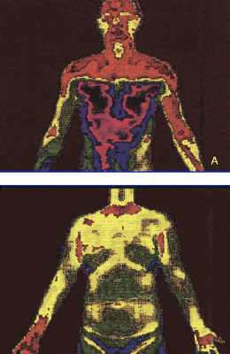
Digital infrared thermal image after T2 sympathicotomy or T3 ramicotomy.

(A) Clear cut change of skin temperature after a T2 sympathicotomy. (B) An even distribution of skin temperature after ramicotomy.
Gossot and colleagues [8] analyzed a group of T2, T3, T4 sympathectomy patients in comparison with a group of patients undergoing a T2, T3, T4 ramicotomy and they reported no statistical difference regarding the incidence of CS between the two groups studied (72.2% and 70.9%). However in terms of the severity of CS (embarrassing, disabling) causing inconveniences to daily life, they reported 27% and 13% incidences in these two groups, respectively. These findings suggest that by preserving the sympathetic trunk, it was possible to reduce the severity of CS.
The preganglionic fibers of the sympathetic nerve to the arm originate mostly from the spinal segments T3–T6 and the postganglionic fibers of the sympathetic nerve to the arm originate from T2 and, to a lesser extent, the T3 ganglia [9]. This implies that the division of preganglionic fibers (rami communicantes) reduces the extent of denervation of the sympathetic nerve as compared with the division of postganglionic fibers (sympathetic trunk) in the treatment of palmar hyperhidrosis.
Sympathectomy or sympathicotomy is one of the procedures used to divide the sympathetic trunk. Sympathicotomy distinctively changes sympathetic nerve distribution in comparison with a ramicotomy. Figure 4A illustrates the clear-cut changes of skin temperature after a T2 sympathicotomy. However the overall sympathetic nerve distribution to the body is not markedly changed after a T3 ramicotomy because a T3 ramicotomy is a procedure that is used to divide one of the preganglionic fibers and to preserve the sympathetic trunk. Figure 4B illustrates an even distribution of skin temperature after T3 ramicotomy.
 http://ats.ctsnetjournals.org/cgi/content/full/78/3/1052#FIG4

Tuesday, April 10, 2012

most surgeons do not have a clear understanding of their short-term outcomes for the majority of procedures they perform

The public would probably be surprised to know that most surgeons do not have a clear understanding of their short-term outcomes for the majority of procedures they perform.

Of even greater concern is the lack of data on long-term outcomes associated with surgical interventions.

Many surgeons argue that they are too busy and do not have the time and resources to conduct this sort of follow-up. This is not entirely without foundation, but it does seem difficult to defend a stance that says “I will continue to work feverishly at the operations I do but not assess how successful my results are”.

Guy Maddern (ASERNIP-s): No excuse for poor surgical outcomes

MJA INSIGHT, 8 August 2011